Discover insights with Inside I-MED
Discover the importance of mammography for women's wellbeing and the benefits of cardiac CT Calcium Scoring for estimating your risk of heart attack in this episode of Inside I-MED!

Discover the importance of mammography for women's wellbeing and the benefits of cardiac CT Calcium Scoring for estimating your risk of heart attack in this episode of Inside I-MED!
View, download and share your images on your phone. Access via a SMS link sent within 48hrs after your scan. Your report will also be visible via this link 7 days after your referring practitioner receives it.
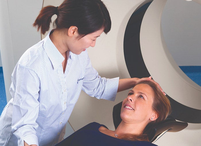
Once our radiologist has finalised your results, I-MED will send a report directly to your referring practitioner. It is important to speak with your health practitioner about your result, to understand what they mean and complete follow up treatment if needed.
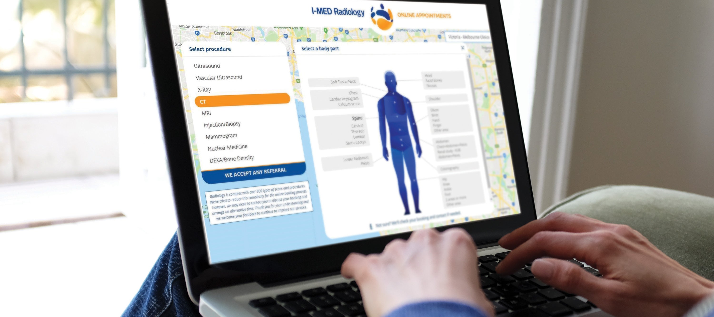
I-MED Radiology clinics offer a range of imaging procedures including MRI, CT, x-ray, ultrasound and nuclear medicine. With the largest team of sub-specialist radiologists, practitioners and patients can be assured of the highest quality imaging and diagnosis.